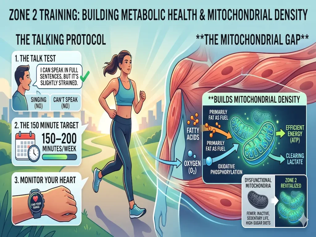
Zone 2 training is performed at an intensity where you are primarily using fat as a fuel source through oxidative phosphorylation in the mitochondria. This builds Mitochondrial Density—the engine of your metabolic health.
The Mitochondrial Gap
Sedentary life and high-sugar diets lead to “mitochondrial dysfunction.” Zone 2 reverses this by forcing your cells to become more efficient at clearing lactate and burning fatty acids.
The Talking Protocol
1. The Talk Test: You should be able to speak in full sentences but sound slightly strained. If you can sing, you’re in Zone 1. If you can’t speak, you’re in Zone 3+.
2. The 150 Minute Target: Aim for 150–200 minutes of Zone 2 per week.
3. Monitor Your Heart: Find your specific Zone 2 range using our [Heart Rate Zones Calculator](/heart-rate-calculator/).
Introduction to the Metabolic Engine
The global rise in metabolic syndrome, type 2 diabetes, and cardiovascular disease has necessitated a profound reevaluation of how therapeutic exercise is prescribed. Central to this evolving paradigm is the concept of Zone 2 training, defined bioenergetically as the exercise intensity at which the body primarily relies on fat as a fuel source through oxidative phosphorylation within the mitochondria.1 By maintaining an intensity that maximizes lipid oxidation while maintaining steady-state blood lactate levels, Zone 2 training directly stimulates the expansion of mitochondrial density—the fundamental engine of human metabolic health.1
Historically, massive volumes of low-intensity training were the exclusive domain of elite endurance athletes, who utilized it to build an aerobic base capable of supporting extreme physiological output.4 However, over the past decade, Zone 2 exercise has transitioned from elite sports science into mainstream preventive medicine. This transition is predicated on the understanding that the root cause of systemic metabolic dysfunction is essentially cellular energetic failure. The modern environment, characterized by chronic physical inactivity and hypercaloric, processed diets, has created what is increasingly termed the “Mitochondrial Gap”—a severe deficit in mitochondrial mass, structural integrity, and oxidative capacity.3
Reversing this gap requires highly specific physiological stimuli. Zone 2 training forces the cells to become radically more efficient at clearing circulating lactate and oxidizing intracellular fatty acids, thereby preventing the lipotoxicity and insulin resistance that characterize metabolic disease.3 Despite its clear biochemical rationale, the widespread promotion of Zone 2 training as a universal panacea for the general public has ignited substantial scientific controversy. Recent exhaustive narrative reviews argue that the physiological adaptations observed in elite athletes who train upwards of forty hours a week cannot be linearly extrapolated to recreational populations exercising for a mere three hours a week.4 This report provides a comprehensive, exhaustive analysis of the biochemical mechanisms underlying mitochondrial dysfunction, the signaling pathways activated by targeted Zone 2 cardiovascular exercise, the revolutionary discoveries regarding the mitochondrial lactate oxidation complex (mLOC), the methodologies for accurately defining and monitoring heart rate zones, and the ongoing academic debate regarding optimal exercise intensity and volume constraints for longevity.
The Mitochondrial Gap: Etiology of Metabolic Dysfunction
To fully appreciate the reparative mechanisms of Zone 2 training, one must first deeply examine the pathological baseline of the modern, industrialized human. The Mitochondrial Gap is not a passive absence of fitness; it is an active state of cellular degeneration driven by the synergistic destruction caused by sedentary behavior and high-sugar diets. This combination systematically dismantles the metabolic engine, leading to profound systemic dysfunction that predates clinical diagnoses of metabolic syndrome by years or even decades.6
The Pathophysiology of Sedentary Lifestyles and Lipotoxicity
In a healthy physiological state, skeletal muscle represents the body’s largest sink for glucose and lipid disposal. This disposal capacity is entirely dependent on the presence of a robust, dense mitochondrial reticulum. However, in a chronically sedentary state, skeletal muscle is deprived of the mechanical and energetic stressors—such as localized ATP depletion, physical membrane stretching, and intracellular calcium fluxes—that are required to maintain mitochondrial quality control.3
Without the stimulus of regular muscular contraction, mitochondrial biogenesis halts, and existing mitochondria undergo structural atrophy.3 This sets the stage for a catastrophic metabolic bottleneck when the individual consumes a modern, lipid-rich, hypercaloric diet. Intramuscular lipid accumulation, a hallmark of insulin resistance and type 2 diabetes, does not result from a primary genetic defect in fatty acid oxidation, as was once widely believed.3 Instead, it stems from a mismatch between lipid uptake and lipid disposal. Fatty acids are continuously taken up into the cell via transport proteins such as FAT/CD36.3 However, because the sedentary individual has an insufficient compensatory increase in mitochondrial mass, these excess fatty acids cannot be oxidized away.3
This state of lipid overload induces intracellular lipotoxicity. Unoxidized lipids are converted into reactive intermediates, such as ceramides and diacylglycerols, which directly interfere with the insulin signaling cascade. These intermediates block the phosphorylation of insulin receptor substrate 1 (IRS-1) and prevent the translocation of GLUT4 transporters to the cell membrane, effectively locking glucose out of the muscle cell and creating systemic hyperinsulinemia.3 This mitochondrial dysfunction is not limited to skeletal muscle; it manifests concurrently in the liver, adipose tissue, heart, vascular endothelium, and pancreatic beta cells, providing a common pathophysiological etiology for chronic diseases ranging from atherosclerosis to neurodegeneration.10 Furthermore, prolonged physical inactivity triggers profound structural degradation down to the skeletal level. The lack of mechanical loading induces osteocyte apoptosis. These dying bone cells release immunostimulatory molecules that instruct local macrophages to produce pro-inflammatory cytokines like TNF-α and IL-6, systemically accelerating inflammation and demonstrating the total-body degradation caused by energetic stagnation.13
Fructose Metabolism, Uric Acid, and the ROS Crisis
While sedentary behavior creates the environment for mitochondrial atrophy, the consumption of the westernized diet—specifically industrialized foods rich in added dietary sugars like sucrose and high-fructose corn syrup—acts as an active agent of mitochondrial destruction.11 The metabolic processing of fructose provides a direct, highly destructive vector for metabolic syndrome that operates completely independently of simple caloric surplus.14
The devastation of fructose occurs primarily in the liver, driven by the unique biochemistry of its processing. When glucose enters a cell, its phosphorylation is strictly regulated by the enzyme phosphofructokinase-1 (PFK-1), which is governed by negative feedback loops; if cellular ATP or citrate levels are high, PFK-1 shuts down, preventing cellular energy overload.15 Fructose bypasses this safety mechanism entirely. Once the polyol pathway or dietary ingestion introduces fructose into the liver, the enzyme fructokinase immediately and irreversibly phosphorylates it into fructose-1-phosphate.15
This unregulated kinase reaction occurs at a rate roughly ten times faster than the phosphorylation of glucose.15 The sheer speed and lack of feedback inhibition drive a massive, acute cellular crisis: the sudden depletion of both intracellular ATP and inorganic phosphate.15 This sudden collapse in the cell’s energy charge activates a specialized survival cascade. The accumulation of AMP triggers the induction of AMP deaminase (AMPD), an enzyme that rapidly shunts the accumulating AMP into the purine degradation pathway.14
The terminal product of this rapid purine catabolism is uric acid.14 A sudden rise in intracellular uric acid is catastrophic for the mitochondria. Uric acid directly induces the generation of mitochondrial reactive oxygen species (ROS), specifically targeting complex I of the electron transport chain.16 While ROS in small, controlled amounts are essential signaling molecules, the massive quantities generated by fructose-induced uric acid accumulation cause severe oxidative damage to mitochondrial DNA, protein aggregations, and lipid peroxidation of the mitochondrial membranes.6
To prevent necrotic cell death under this extreme oxidative stress, the cell is forced to initiate mitophagy—the targeted destruction and removal of its own damaged mitochondria.6 This aggressively reduces the total mitochondrial number, further decreasing the cell’s ability to oxidize substrates and severely aggravating lipid accumulation.6
Simultaneously, the activation of AMPD downregulates AMP-activated protein kinase (AMPK).15 Because AMPK is the master regulator responsible for fat oxidation and mitochondrial biogenesis, its deactivation is the final blow to metabolic health. With AMPK suppressed, acetyl-CoA carboxylase 2 is deactivated, and carbohydrate response element-binding protein (ChREBP) is simultaneously upregulated.15 This biochemical combination forcibly drives de novo lipogenesis, directly leading to Non-Alcoholic Fatty Liver Disease (NAFLD).15 The systemic consequences are profound. Pediatric obesity and NAFLD have surged in parallel with beverage consumption, becoming the major cause of chronic liver disease in childhood.17 Fructose ingestion further disrupts tight junctions in the intestine, increasing gut permeability and allowing endotoxins to enter the portal vein, which heavily triggers fatty liver formation and systemic subclinical atherosclerotic damage even at a very early age.17 Even more alarming, the diet-induced accumulation of advanced glycation end-products (AGEs) and the consumption of high-sugar diets by mothers during gestation epigenetically program their offspring with an exponentially increased risk of developing type 2 diabetes later in life, perpetuating the disease across generations.11
Physiological Mechanisms of Zone 2 Training
To combat the Mitochondrial Gap and reverse the lipotoxic effects of the modern diet, an intervention must simultaneously upregulate lipid oxidation and stimulate the structural synthesis of new mitochondria. Zone 2 training achieves precisely this by maintaining cardiovascular exercise at an intensity where the muscular energy demand is met almost exclusively by oxidative phosphorylation, using fatty acids as the primary substrate.1
Biochemical Signaling and Mitochondrial Biogenesis
The structural expansion of the mitochondrial reticulum is not a generalized response to exertion; it is a highly specific architectural adaptation driven by precise biochemical signaling networks. During sustained, low-to-moderate intensity Zone 2 exercise, the continuous hydrolysis of ATP by contracting skeletal muscles results in a modest but persistent accumulation of AMP and ADP.2
This subtle shift in the intracellular energy charge acts as the primary mechanical and metabolic trigger. The elevated AMP levels are sensed by AMP-activated protein kinase (AMPK), often described as the master energy sensor of the cell.12 Once activated, AMPK initiates a multi-tiered signaling cascade designed to restore the cellular energy balance by increasing the cell’s capacity for oxidative metabolism.12 AMPK directly phosphorylates and activates a host of downstream targets, but its most critical interaction is with SIRT1 (Sirtuin 1), an NAD+-dependent deacetylase.12
AMPK and SIRT1 operate in a highly synchronized partnership to control energy expenditure. Together, they target the peroxisome proliferator-activated receptor-gamma coactivator 1-alpha (PGC-1α).12 PGC-1α is the supreme master transcriptional coactivator responsible for driving mitochondrial biogenesis.3 AMPK phosphorylates PGC-1α, while SIRT1 deacetylates it, leading to its full activation.12
Once activated, PGC-1α translocates into the cell nucleus, where it radically alters gene expression. It co-activates nuclear respiratory factors (NRF-1 and NRF-2) and estrogen-related receptor alpha (ERRα), which then drive the transcription of nuclear-encoded mitochondrial proteins, while simultaneously stimulating mitochondrial DNA replication.18 This process physically builds new mitochondria and increases the cristae density within existing ones.18
Parallel to the AMPK pathway, the continuous mechanical stress and regular intracellular calcium fluxes inherent in the repetitive muscle contractions of Zone 2 cycling, running, or swimming activate calcium/calmodulin-dependent protein kinase II (CaMKII).18 CaMKII provides a secondary, robust pathway to PGC-1α activation, ensuring that the mechanotransduction of physical movement is directly translated into biochemical adaptation.18 Additionally, AMPK regulates mitochondrial dynamics, managing the delicate balance of mitochondrial fission and fusion to maintain a healthy reticular network, while simultaneously initiating autophagy flux to selectively degrade and recycle old, dysfunctional mitochondria.12
Fat Oxidation Capacity and Metabolic Flexibility
The immediate functional outcome of this PGC-1α-driven mitochondrial biogenesis is a massive upgrade in the cell’s capacity to transport and oxidize fatty acids. Zone 2 training systematically upregulates the expression of key lipid transport and oxidative enzymes, including FAT/CD36 (for transporting fatty acids across the sarcolemma), FABP3, carnitine palmitoyltransferase I (CPT1B, which shuttles long-chain fatty acids into the mitochondria), and β-HAD (a critical enzyme in the beta-oxidation cycle).1
By expanding the mitochondrial volume and enzymatic machinery, Zone 2 training effectively eliminates the lipid disposal bottleneck that causes insulin resistance.3 As the peak fat oxidation rate rises over 8 to 12 weeks of consistent low-intensity volume, the individual’s physiology undergoes a profound shift.1 They develop an immense capacity to burn fat not just at rest, but at increasingly higher absolute exercise intensities.1
This enhanced fat oxidation crucially spares the body’s limited glycogen reserves.1 Because Zone 2 relies heavily on lipids, it causes minimal glycogen depletion, allowing athletes to delay the onset of fatigue and reducing the total volume of carbohydrates burned at any given endurance pace.1 The ultimate expression of this adaptation is “metabolic flexibility”—the capacity of the human body to seamlessly transition back and forth between lipid and carbohydrate utilization based strictly on the acute physiological demand.21 Elite endurance athletes demonstrate legendary metabolic flexibility, maintaining peak fat oxidation at power outputs that would force a metabolically inflexible, sedentary individual to rely entirely on anaerobic glycolysis, leading to rapid exhaustion and systemic acidification.22
The Lactate Paradigm and the Mitochondrial Lactate Oxidation Complex
Perhaps the most revolutionary shift in modern exercise physiology, directly bearing on the efficacy of Zone 2 training, involves the complete reclassification of lactate. For nearly a century, sports scientists, physicians, and coaches erroneously viewed lactate (lactic acid) as a toxic, dead-end metabolic waste product that caused muscle fatigue, delayed onset muscle soreness, and cellular acidosis.24 The foundational work of Dr. George Brooks fundamentally destroyed this dogma, revealing lactate to be arguably the most dynamic energy substrate in the human body.26
The Cell-Cell Lactate Shuttle Theory
Introduced in 1984 by George Brooks, the Lactate Shuttle Hypothesis posited that lactate is not a waste product, but a highly mobile fuel source, the body’s primary gluconeogenic precursor, and a potent signaling molecule with autocrine, paracrine, and endocrine functions.25
During exercise, glycolysis breaks down glucose to produce pyruvate. To regenerate NAD+ so that glycolysis can continue operating at high speeds, pyruvate is rapidly reduced to lactate.28 The “Cell-Cell Lactate Shuttle” describes the process by which lactate is produced in one tissue bed (primarily fast-twitch, highly glycolytic Type II muscle fibers) and immediately transported through the bloodstream to better-oxygenated domains where it can be consumed as fuel.8
Lactate is heavily favored as an oxidative fuel by the working heart muscle, the liver, the kidneys, and even the brain.8 The brain’s neurons are heavily dependent on lactate for long-term memory formation and high-level cognitive function during physiological stress.8 The transport of lactate across cell membranes is governed by a family of monocarboxylate transporters (MCTs). Glycolytic fast-twitch fibers possess a high density of MCT4 transporters, which are specialized to export lactate out of the cell.8 Conversely, highly oxidative slow-twitch Type I fibers, heart muscle, and neuronal tissues are rich in MCT1 transporters, which act as vacuums to pull lactate inside the cell for utilization.8
The defining characteristic of highly trained endurance athletes is not that they produce less lactate; rather, they possess an extraordinary, enhanced lactate clearance capacity.8 Their robust mitochondrial networks clear lactate directly within the producing muscle itself or shuttle it immediately to adjacent oxidative fibers, preventing systemic blood lactate levels from rising even at blistering speeds.8 The importance of lactate shuttling extends far beyond sports performance. Dysfunctional lactate shuttling is now deeply implicated in the etiology of cancer, chronic inflammation, and severe metabolic inflexibility.25
The Intracellular Shuttle and the mLOC
In 1998, Brooks extended his hypothesis to include the “Intracellular Lactate Shuttle,” proposing that lactate produced in the cytosol could be oxidized directly by the mitochondria within the very same cell.27 This concept faced intense resistance from the biological establishment, which rigidly adhered to the dogma that only pyruvate could cross the inner mitochondrial membrane.27 However, modern molecular biology, utilizing confocal laser scanning microscopy (CLSM), 3D in situ proximity ligation, and immunoprecipitation, has definitively proven Brooks correct.32
The mechanism enabling this intracellular shuttling is the Mitochondrial Lactate Oxidation Complex (mLOC), an incredibly sophisticated structural and functional apparatus physically embedded in the mitochondrial inner membrane.28 The mLOC has been definitively mapped in skeletal muscle, heart tissue, and notably, within the mitochondria of neurons in the cortex and hippocampus, proving that cerebral neurons utilize this complex to oxidize lactate delivered by adjacent astrocytes.32
Based on groundbreaking 2024 and 2025 research published in the American Journal of Physiology-Endocrinology and Metabolism, the structural composition of the mLOC is currently understood to consist of five key interdependent members 28:
| mLOC Protein Component | Functional Role in Carbohydrate Carbon Disposal |
| mMCT1 (Mitochondrial Monocarboxylate Transporter 1) | Embedded in the inner mitochondrial membrane, this transporter facilitates the direct, rapid import of lactate and pyruvate from the cytosol into the mitochondrial matrix. |
| CD147 (Basigin) | An essential scaffolding glycoprotein that acts as an anchoring protein, maintaining the structural integrity and precise positioning of mMCT1 within the complex. |
| mLDH (Mitochondrial Lactate Dehydrogenase) | Positioned primarily within the mitochondrial cristae. Once lactate enters via mMCT1, mLDH catalyzes the endergonic conversion of lactate back into pyruvate inside the mitochondrion. |
| COx (Cytochrome c Oxidase) | Known as Complex IV of the electron transport chain. The exergonic redox changes occurring during electron transport in COx are tightly coupled to mLDH, driving the energetically demanding oxidation of lactate. |
| mPC (Mitochondrial Pyruvate Carrier) | The most recently confirmed member (2024/2025). Works synergistically within the complex to manage the lower end of the concentration gradient, ensuring seamless final disposal of pyruvate into the TCA cycle. |
Structural data compiled from Leija et al. (2025).28
This complex fundamentally rewrites the final pathway of carbohydrate oxidation.28 Because of the extreme efficiency of the mLOC, mitochondrial lactate oxidation actually dominates over pyruvate oxidation in healthy, active tissues.24 However, if lactate accumulates and cannot be cleared—due to a lack of MCT1 transporters or mitochondrial density—the elevated intracellular lactate binds to G protein-coupled receptors (HCAR1) and actively blocks lipolysis and mitochondrial fatty acid uptake.38 Thus, poor lactate clearance actively shuts down fat burning.
Zone 2 training is the supreme intervention for optimizing the mLOC. By performing long-duration exercise precisely at the intensity where lactate production is perfectly matched by lactate clearance, the body is forced to adapt by radically increasing the expression of MCT1.30 Zone 2 training facilitates the massive insertion of MCT1 transporters into both the sarcolemmal and mitochondrial membranes, directly augmenting the cell’s ability to pull lactate out of the blood and burn it in the mitochondria alongside fatty acids.30
Establishing and Validating Training Zones
Because the biochemical cascades governing fat oxidation and lactate shuttling are highly intensity-dependent, the physiological benefits of Zone 2 training vanish if the exercise intensity drifts too high. Crossing the critical physiological threshold shifts the body’s substrate reliance from lipids to carbohydrates, causing an exponential rise in blood lactate that overwhelms the mLOC and shuts down the specific AMPK/PGC-1α signaling pathways targeted by aerobic base training.40 Consequently, precise methodologies for defining heart rate zones and physiological thresholds are absolutely critical.
Laboratory vs. Field Testing Methodologies
The scientific gold standard for establishing individual training zones is rigorous laboratory testing.40 These assessments utilize medical-grade cardiopulmonary metabolic carts and direct blood sampling to remove all environmental noise and subjective estimation.40
During a laboratory test, the athlete exercises on a treadmill or a stationary bike (such as a Wahoo Kickr) while wearing a specialized mask that captures expired gases.40 The intensity is progressively increased in staged intervals. The metabolic cart analyzes breath-by-breath gas exchange to quantify exact oxygen uptake () and carbon dioxide production (
), while researchers simultaneously collect capillary blood from the earlobe or fingertip to plot the precise lactate accumulation curve.41
This allows physiologists to pinpoint exact inflection points. The first clear rise in breathing relative to oxygen use marks Ventilatory Threshold 1 (VT1), the ceiling of Zone 2.40 The point at which lactate begins accumulating faster than the mLOC can clear it marks the Lactate Threshold (LT).42 Furthermore, laboratory testing quantifies the exact Respiratory Exchange Ratio (RER), allowing practitioners to calculate practical carbohydrate vs. fat oxidation rates per hour at target intensities.40
Conversely, field testing estimates thresholds using mathematical modeling or maximal performance efforts.40 While infinitely more accessible, field tests are highly vulnerable to environmental factors like heat, wind, and course profiles, and their accuracy relies entirely on the athlete’s psychological motivation and pacing strategy.40
The table below contrasts the distinct approaches and outcomes of Laboratory versus Field Testing for establishing thresholds:
| Parameter | Laboratory Testing (Metabolic Cart & Direct Blood Sampling) | Field Testing (Time Trials & Mathematical Modeling) |
| Testing Environment | Highly controlled clinical environment; eliminates external variables (wind, terrain, temperature) ensuring repeatable protocols. | Outdoors or on personal equipment; highly susceptible to environmental “noise” which can skew data by 3% to 8%. |
| Measurement Mechanism | Direct measurement of breath-by-breath gas exchange (VO2 max) and chemical blood lactate kinetics via progressive stages. | Retrospective estimation using external markers (average pace/power over 30-60 mins) or Critical Power curve modeling. |
| Threshold Accuracy | Objectively pinpoints exact physiological shifts: Ventilatory Threshold 1 (VT1), VT2, and true Lactate Threshold (LT). | Yields a mathematically estimated Functional Threshold Power/Pace and an estimated Lactate Threshold Heart Rate (LTHR). |
| Substrate & Fueling Insights | Maps exact fat-burning efficiency, fat vs. carbohydrate oxidation ratios (RER), dictating precise exogenous fueling requirements for race day. | Provides zero metabolic measurements or substrate utilization insights; offers no data on fat vs. carbohydrate usage. |
| Execution Dependency | Relies on forced progressive stages; requires no pacing expertise from the athlete to yield accurate results. | Heavily dependent on the athlete’s pacing experience; poor pacing or lack of maximal motivation renders the data entirely unreliable. |
Comparison based on human performance testing standards detailed in.40
If laboratory testing is unavailable, athletes commonly utilize Joe Friel’s field testing protocol to establish their Lactate Threshold Heart Rate (LTHR). This involves executing a solo, 30-minute maximal time trial, taking the average heart rate of the final 20 minutes as the LTHR.40 Crucially, zones must be calculated specific to the modality, as weight-bearing activities elicit different cardiovascular responses. The Friel zone mapping is distinct for cycling and running 43:
| Training Zone | Running Prescription (% of LTHR) | Cycling Prescription (% of LTHR) |
| Zone 1 (Active Recovery) | Less than 85% | Less than 81% |
| Zone 2 (Aerobic Endurance) | 85% to 89% | 81% to 89% |
| Zone 3 (Tempo) | 90% to 94% | 90% to 93% |
| Zone 4 (Sub-Threshold) | 95% to 99% | 94% to 99% |
| Zone 5a/b/c (Maximal) | > 100% | > 100% |
Friel’s established endurance zones based on LTHR.43
Validating Maximum Heart Rate Equations
For individuals utilizing commercial wearables that rely on a percentage of Maximum Heart Rate (), the underlying mathematical formula is critical. For decades, the global standard was the ubiquitous Fox equation:
.44 However, this equation was never rigorously validated in large epidemiological samples containing older adults, leading to severe underestimations of maximum heart rate in older populations, which subsequently caused under-prescription of exercise intensity.45
To rectify this, a landmark meta-analysis was conducted by Tanaka, Monahan, and Seals (2001), reviewing 351 studies encompassing 18,712 subjects across 492 groups.45 They derived a new, highly accurate generalized equation, which was then cross-validated in a rigorously controlled laboratory trial of 514 healthy subjects.45 The resultant Tanaka Formula:

The validation trials demonstrated that the regression line for this formula was virtually identical across diverse populations, proving that is predicted to a large extent by age alone and is completely independent of gender or habitual physical activity status.45 Comparative analyses against other prevalent formulas continuously validate the superiority of the Tanaka equation.
The table below outlines the evolution and variations of maximum heart rate prediction equations historically proposed in the literature 44:
| Author / Equation Origin | Heart Rate Maximum Prediction Formula | Primary Application / Population Notes |
| Fox (Classic) | Widely used historically; proven to severely underestimate max HR in older adults. | |
| Tanaka (2001) | The modern gold standard; validated across 18,000+ subjects; independent of gender and fitness. | |
| Gellish | Nearly identical mathematically to Tanaka; frequently applied in clinical testing. | |
| Gulati | Specifically derived and validated for female populations to account for minor physiological deviations. | |
| Arena | Refined mathematical modeling; closely tracks the Tanaka and Gellish meta-analyses. | |
| Astrand | Older classical model; tends to overestimate HR in younger populations compared to modern data. | |
| Nes (2013) | A more recent formula heavily applied in Scandinavian epidemiological research. |
Summary of HR prediction equations from cardiological validation studies.44
For predicting observed values of , the Tanaka formula consistently calculates the smallest signed and unsigned residuals from the difference between observed and predicted values, confirming it as the most accurate generalized tool for setting the ceiling on Zone 2 calculations.45
The Controversy: “Much Ado About Zone 2”
The flawless biochemical logic underlying lipid oxidation and lactate shuttling has propelled Zone 2 training into the zenith of popular health culture. Podcasts, social media influencers, and longevity physicians have aggressively touted Zone 2 as the absolute optimal training intensity for improving mitochondrial capacity, supporting metabolic health, and driving chronic disease prevention.4
However, a fierce counter-movement within the academic community has emerged, challenging the universal applicability of this doctrine. A critical 2025 narrative review published in Sports Medicine, titled “Much Ado About Zone 2,” fundamentally interrogates the efficacy of low-intensity volume when prescribed to the general, time-poor public.4
The Illusion of the Elite Training Model
The broad endorsement of Zone 2 training stems largely from observational data derived from elite endurance athletes.4 Cyclists, marathoners, and triathletes possess staggering mitochondrial and fatty acid oxidative capacities, and a retrospective analysis of their training logs reveals a polarized model where roughly 80% of their training volume is performed at a low intensity (Zone 2).4 The logical leap made by popular media was that the low intensity itself was the magic bullet producing the exceptional mitochondrial health.5
The “Much Ado” review exposes the fundamental flaw in this extrapolation: absolute volume.5 Elite athletes train between 30 and 40 hours per week. While 80% of their volume is low-intensity, the remaining 20% of their high-intensity interval training still equates to 6 to 8 hours of brutal, vigorous work per week.5 The extraordinary adaptations observed in these athletes may be driven primarily by the sheer magnitude of their high-intensity volume, rather than the low-intensity work per se.4
When clinical researchers, such as PhD candidate Kristi Storoschuk, actively search the literature for studies explicitly examining physiological outcomes driven exclusively by Zone 2 training as it is commonly characterized, the substantive evidence is surprisingly scarce.4 When low-intensity continuous exercise is compared directly against high-intensity interval training (HIIT) in volume-matched studies, higher intensity consistently wins.5 A comprehensive 2018 meta-analysis by Granada and colleagues demonstrated that mitochondrial respiratory capacity and structural adaptations respond most robustly when exercise is performed at or above 65% to 90% of a subject’s maximum work rate.4 Sprint interval training repeatedly proves more effective at surging PGC-1α and driving rapid mitochondrial biogenesis than isocaloric low-intensity work.4
The Volume Constraint and the Danger of Misprescription
The danger of the Zone 2 narrative lies in its application to the general public, who typically adhere to physical activity guidelines recommending 150 to 300 minutes of total exercise per week.2 Within this severely constrained time budget, the absolute volume of Zone 2 training is mathematically insufficient to mimic the adaptations of the elite model.50
Zone 2 is physiologically defined by keeping blood lactate concentrations tightly controlled below 2.0 millimoles per liter.5 For an average individual, exercising below this 2.0 mmol/L threshold generates a signaling amplitude that simply falls below the adaptive threshold required to reliably rescue their physiology from established mitochondrial dysfunction.5 The “Much Ado” authors argue that advising a metabolically compromised individual to forgo higher exercise intensities in favor of purely comfortable, sub-threshold Zone 2 work may drastically limit the health benefits of their limited exercise window.4
Because Zone 2 falls below the moderate-to-vigorous intensity range needed to force massive cardiovascular remodeling, the current evidence does not support Zone 2 as the optimal standalone intensity for the general public.4 The narrative review concludes that prioritizing high-intensity exercise (> Zone 2) is strictly critical to maximizing cardiometabolic health benefits, particularly in the context of lower training volumes typical of modern lifestyles.4
Operational Protocols and Practical Implementation
Synthesizing the undeniable cellular benefits of building the mitochondrial lactate oxidation complex with the clinical realities of the volume constraint requires a highly structured, individualized implementation protocol.
The Talking Protocol
To effectively lock into Zone 2 without the expense of a laboratory metabolic cart or the pain of a time trial, practitioners universally recommend the validated “Talking Protocol.” This relies on the physiological realities of the ventilatory thresholds (VT1 and VT2). As exercise intensity rises, the accumulating from glycolytic buffering forces the respiratory rate to increase, which directly impedes speech.40
The precise implementation of the Talk Test dictates the following rules for execution:
- The Talk Test: During continuous exertion, the individual must attempt to speak aloud in full, complete sentences. To be squarely in Zone 2, the speech should be possible, but the voice must sound slightly strained, requiring conscious breath management between sentences.
- If you can sing effortlessly, the intensity is far too low; you are in Zone 1, failing to recruit sufficient motor units to stress the mitochondrial network.
- If you can’t speak in full sentences, and are reduced to gasping short phrases or single words, you have crossed Ventilatory Threshold 2 (VT2) into Zone 3 or higher. At this point, glycolytic flux has overwhelmed the mLOC’s clearing capacity, lactate is pooling, and you are no longer prioritizing fat oxidation.
- The 150 Minute Target: To achieve baseline structural shifts in mitochondrial density and upregulate MCT1 expression, individuals must aim for an absolute minimum of 150 to 200 minutes of precise Zone 2 training per week.50 For time-poor executives, this translates to three or four dedicated sessions ranging from 45 to 90 minutes each.50
- Monitor Your Heart: To ensure compliance across changing terrain and daily fatigue levels, individuals must anchor their perceived exertion to data. Practitioners instruct individuals to find their specific Zone 2 range using our Heart Rate Zones Calculator, which applies formulas like the Tanaka equation to provide an objective ceiling on daily efforts.
Genetic Variability and Overtraining
Finally, the application of these protocols must respect individual genetic variability. Variations in the PPARGC1A gene significantly alter how rapidly an individual responds to aerobic volume.50 “High responders” may see rapid expansions in their max and lactate clearing capacities within weeks, while “low responders” require significantly more volume or a longer timeline spanning months to achieve identical gains.50
Baseline fitness cannot be ignored. For an individual who has been completely sedentary for years, the initial mitochondrial atrophy is so severe that a brisk walk may push their blood lactate above 2.0 mmol/L, placing them physiologically in Zone 3 despite the low mechanical output.50 In these cases, building the base requires extreme patience and walking protocols until the mLOC architecture is established.50 Furthermore, while elites manage 40 hours a week, recreational athletes must understand that more is not unilaterally better; excessive volume layered onto a stressful lifestyle without adequate sleep leads directly to overtraining, immune suppression, and injury, collapsing the very metabolic architecture the exercise sought to build.2
Conclusion
The architecture of optimal human metabolic health is inextricably linked to the physical density and functional capacity of the mitochondrial reticulum. The modern environment—defined by the cessation of physical movement and the chronic consumption of unregulated substrates like dietary fructose—initiates a devastating pathological cascade. ATP depletion, the induction of AMP deaminase, uric acid generation, and the suppression of AMPK systematically dismantle the mitochondria, leading directly to lipotoxicity, insulin resistance, and the generational propagation of metabolic syndrome.
Zone 2 training offers a profound, mechanically elegant remedy to this Mitochondrial Gap. By enforcing a precise bioenergetic environment characterized by low glycolytic stress and maximum rates of lipid oxidation, it directly targets the AMPK and SIRT1 pathways to unleash PGC-1α, driving explosive mitochondrial biogenesis. It optimizes the remarkable machinery of the newly mapped mitochondrial lactate oxidation complex (mLOC), allowing the seamless transport of lactate via mMCT1 and mPC, and its subsequent conversion and oxidation by mLDH and Cytochrome c Oxidase. This capability transforms lactate from a fatiguing waste product into a primary fuel source, restoring supreme metabolic flexibility.
Yet, translating this physiological ideal into practical health interventions requires a rejection of dogma. The uncritical promotion of Zone 2 training as the sole requirement for health ignores the rigorous volume constraints of the general public. While Zone 2 builds the essential metabolic engine, relying on it exclusively within a 150-minute weekly budget fails to provide the high-amplitude signaling required for maximum cardiometabolic adaptation. Accurate prescription utilizing the Talking Protocol, the Tanaka formula, and the Heart Rate Zones Calculator ensures the foundation is built correctly. Ultimately, however, defending against chronic metabolic disease requires the structural, lipid-oxidizing foundation of Zone 2 exercise to be intelligently augmented by the potent, adaptive stressors of high-intensity effort.
Works cited
- Does Zone 2 Improve Fat Oxidation? — What the Science Shows | Roadman Cycling, accessed June 4, 2026, https://roadmancycling.com/answers/zone-2-fat-oxidation
- Do We Really Need Zone 2 Exercise for Mitochondrial and …, accessed June 4, 2026, https://brokenscience.org/do-we-really-need-zone-2-exercise-for-mitochondrial-and-metabolic-health/
- PGC-1α regulation by exercise training and its influences on muscle function and insulin sensitivity – PMC, accessed June 4, 2026, https://pmc.ncbi.nlm.nih.gov/articles/PMC2928513/
- A Narrative Review Assessing the Efficacy of Zone 2 Training for Improving Mitochondrial Capacity and Cardiorespiratory Fitness in the General Population, accessed June 4, 2026, https://www.fisiologiadelejercicio.com/wp-content/uploads/2025/06/Much-Ado-About-Zone-2.pdf
- A Mitochondria Researcher Went Looking for Evidence to Support Zone 2. Here Is What She Found. | Healthspan, accessed June 4, 2026, https://www.gethealthspan.com/research/article/zone-2-training-longevity-evidence
- Mitochondrial dysfunction and insulin resistance: an update – PMC, accessed June 4, 2026, https://pmc.ncbi.nlm.nih.gov/articles/PMC4261703/
- Mitochondrial (Dys)function and Insulin Resistance: From Pathophysiological Molecular Mechanisms to the Impact of Diet – PMC, accessed June 4, 2026, https://pmc.ncbi.nlm.nih.gov/articles/PMC6510277/
- The Lactate Shuttle Theory – Roberto Vukovic, accessed June 4, 2026, https://robertovukovic.com/lactate-shuttling/
- Much Ado About Zone 2: A Narrative Review Assessing the Efficacy of Zone 2 Training for Improving Mitochondrial Capacity and Cardiorespiratory Fitness in the General Population | springermedicine.com, accessed June 4, 2026, https://www.springermedicine.com/much-ado-about-zone-2-a-narrative-review-assessing-the-efficacy-/51145116
- Role of Mitochondrial Dysfunction in Insulin Resistance | Circulation Research, accessed June 4, 2026, https://www.ahajournals.org/doi/10.1161/circresaha.107.165472
- The Bitter Side of Sugar Consumption: A Mitochondrial Perspective on Diabetes Development – MDPI, accessed June 4, 2026, https://www.mdpi.com/2673-4540/3/4/44
- Institutional Repository – Research Portal Dépôt Institutionnel – Portail de la Recherche – researchportal.unamur.be – Sign in to Université de Namur, accessed June 4, 2026, https://pure.unamur.be/ws/portalfiles/portal/62327161/2022_JuszcakF_these.pdf
- Molecular Mechanisms of Obesity-Induced Osteoporosis and Muscle Atrophy – PMC – NIH, accessed June 4, 2026, https://pmc.ncbi.nlm.nih.gov/articles/PMC5040721/
- Morin Alleviates Fructose-Driven Disturbance of Podocyte Mitochondrial Energy Metabolism by Inhibiting Adenosine 5′-Monophosphate Deaminase Activity to Improve Glomerular Injury – PMC, accessed June 4, 2026, https://pmc.ncbi.nlm.nih.gov/articles/PMC12736346/
- Aldose reductase, fructose and fat production in the liver – Portland Press, accessed June 4, 2026, https://portlandpress.com/biochemj/article/482/05/295/235710/Aldose-reductase-fructose-and-fat-production-in
- Newly discovered mechanism of mitochondrial dysfunction in obesity may drive insulin resistance and type 2 diabetes, accessed June 4, 2026, https://hsph.harvard.edu/news/newly-discovered-mechanism-of-mitochondrial-dysfunction-in-obesity-may-drive-insulin-resistance-and-type-2-diabetes/
- Fructose and Uric Acid: Major Mediators of Cardiovascular Disease Risk Starting at Pediatric Age – PMC, accessed June 4, 2026, https://pmc.ncbi.nlm.nih.gov/articles/PMC7352635/
- Unravelling the mechanisms regulating muscle mitochondrial biogenesis | Request PDF, accessed June 4, 2026, https://www.researchgate.net/publication/305716682_Unravelling_the_mechanisms_regulating_muscle_mitochondrial_biogenesis
- Can exercise mitigate the negative metabolic effects associated with sleep loss? – VU Research Repository, accessed June 4, 2026, https://vuir.vu.edu.au/40011/1/SANER%20Nicholas-thesis_nosignature.pdf
- Exercise Training Induced Improvement in Skeletal Muscle PGC-1α Mediated Fat Metabolism is Independent of Dietary Glycemic Index – PMC, accessed June 4, 2026, https://pmc.ncbi.nlm.nih.gov/articles/PMC5373498/
- Lactate: The Key to Metabolic Health, Mitochondria, and Longevity | Dr Iñigo San Millán, accessed June 4, 2026, https://theproof.com/lactate-the-key-to-metabolic-health-mitochondria-and-longevity-dr-inigo-san-millan/
- Inigo San Millan, PhD | Profiles | School of Medicine | University of Colorado – CU Anschutz, accessed June 4, 2026, https://som.cuanschutz.edu/Profiles/Faculty/Profile/19887
- Metabolic and Cellular Differences Between Sedentary and Active Individuals at Rest and During Exercise | bioRxiv, accessed June 4, 2026, https://www.biorxiv.org/content/10.1101/2024.08.19.608601v1.full
- Lactate: The Unsung Hero of Endurance Metabolism | Uphill Athlete, accessed June 4, 2026, https://uphillathlete.com/aerobic-training/lactate-the-unsung-hero-of-endurance-metabolism/
- The tortuous path of lactate shuttle discovery: From cinders and boards to the lab and ICU, accessed June 4, 2026, https://pmc.ncbi.nlm.nih.gov/articles/PMC7498672/
- Lactate metabolism: a new paradigm for the third millennium – PMC, accessed June 4, 2026, https://pmc.ncbi.nlm.nih.gov/articles/PMC1664920/
- Tracing the lactate shuttle to the mitochondrial reticulum – PMC – NIH, accessed June 4, 2026, https://pmc.ncbi.nlm.nih.gov/articles/PMC9534995/
- The mitochondrial lactate oxidation complex: endpoint for carbohydrate carbon disposal | American Journal of Physiology-Endocrinology and Metabolism, accessed June 4, 2026, https://journals.physiology.org/doi/abs/10.1152/ajpendo.00306.2024
- The mitochondrial lactate oxidation complex: endpoint for carbohydrate carbon disposal, accessed June 4, 2026, https://pmc.ncbi.nlm.nih.gov/articles/PMC12145959/
- Science, New Interval Training, Lactate Shuttle, MCTs, George Brooks, accessed June 4, 2026, https://www.newintervaltraining.com/the-science.php
- Iñigo San Millán Physiology and Metabolism in Health & Disease University of Colorado – ResearchGate, accessed June 4, 2026, https://www.researchgate.net/profile/Inigo-San-Millan
- Evidence for the Mitochondrial Lactate Oxidation Complex in Rat Neurons: Demonstration of an Essential Component of Brain Lactate Shuttles – eScholarship.org, accessed June 4, 2026, https://escholarship.org/uc/item/2075h0v7
- The mitochondrial lactate oxidation complex: endpoint for carbohydrate carbon disposal | American Journal of Physiology-Endocrinology and Metabolism, accessed June 4, 2026, https://journals.physiology.org/doi/prev/20241223-aop/abs/10.1152/ajpendo.00306.2024
- Evidence for the Mitochondrial Lactate Oxidation Complex in Rat Neurons: Demonstration of an Essential Component of Brain Lactate Shuttles – PMC, accessed June 4, 2026, https://pmc.ncbi.nlm.nih.gov/articles/PMC2488371/
- Prof. Dr. George Brooks | Author – SciProfiles, accessed June 4, 2026, https://sciprofiles.com/profile/1159305
- The mitochondrial lactate oxidation complex: endpoint for carbohydrate carbon disposal, accessed June 4, 2026, https://pubmed.ncbi.nlm.nih.gov/39714986/
- Lactate Metabolism and The Mitochondrial Reticulum: Enteric and Systemic Shuttles, Oxidation, and Aging – UC Berkeley, accessed June 4, 2026, https://escholarship.org/content/qt3km7s2w8/qt3km7s2w8.pdf
- The mitochondrial lactate oxidation complex: endpoint for carbohydrate carbon disposal – eScholarship.org, accessed June 4, 2026, https://escholarship.org/content/qt6md7m998/qt6md7m998.pdf
- Lactate transported by MCT1 plays an active role in promoting mitochondrial biogenesis and enhancing TCA flux in skeletal muscle – PMC, accessed June 4, 2026, https://pmc.ncbi.nlm.nih.gov/articles/PMC11204292/
- Find Your Threshold: Field Tests Vs. Lab Testing For Anaerobic And …, accessed June 4, 2026, https://metatec.org/find-your-threshold-field-tests-vs-lab-testing-for-anaerobic-and-lactatethreshold/
- Training Zones Determination – Human Performance Clinical Research Laboratory, accessed June 4, 2026, https://www.chhs.colostate.edu/hes-hpcrl/performance-health-analysis/training-zones/
- Understanding Heart Rate Training Zones, V02 Max, & Lactate Threshold, accessed June 4, 2026, https://bandanatraining.com/heart-rate-training-zones/
- Joe Friel’s Quick Guide to Setting Zones | – TrainingPeaks, accessed June 4, 2026, https://www.trainingpeaks.com/learn/articles/joe-friel-s-quick-guide-to-setting-zones/
- Accuracy of Commonly Used Age-Predicted Maximal Heart Rate Equations – PMC, accessed June 4, 2026, https://pmc.ncbi.nlm.nih.gov/articles/PMC7523886/
- Age-predicted maximal heart rate revisited – PubMed – NIH, accessed June 4, 2026, https://pubmed.ncbi.nlm.nih.gov/11153730/
- HR Max Prediction Based on Age, Body Composition, Fitness Level, Testing Modality and Sex in Physically Active Population – Frontiers, accessed June 4, 2026, https://www.frontiersin.org/journals/physiology/articles/10.3389/fphys.2021.695950/full
- Validation of Maximal Heart Rate Prediction Equations Based on Sex and Physical Activity Status – PMC, accessed June 4, 2026, https://pmc.ncbi.nlm.nih.gov/articles/PMC4831892/
- Much Ado About Zone 2: A Narrative Review Assessing the Efficacy of Zone 2 Training for Improving Mitochondrial Capacity and Cardiorespiratory Fitness in the General Population – PubMed, accessed June 4, 2026, https://pubmed.ncbi.nlm.nih.gov/40560504/
- Much Ado About Zone 2: A Narrative Review Assessing the Efficacy of Zone 2 Training for Improving Mitochondrial Capacity and Cardiorespiratory Fitness in the General Population : r/PeterAttia – Reddit, accessed June 4, 2026, https://www.reddit.com/r/PeterAttia/comments/1mdv976/much_ado_about_zone_2_a_narrative_review/
- Zone 2 Cardio and Longevity – Superpower, accessed June 4, 2026, https://superpower.com/guides/zone-2-cardio-and-longevity
- Normal Versus Chronic Adaptations to Aerobic Exercise – StatPearls – NCBI Bookshelf, accessed June 4, 2026, https://www.ncbi.nlm.nih.gov/books/NBK572066/
Zone 2 Training for Executives: Why 180 Minutes a Week | Wellness, accessed June 4, 2026, https://wellnesselitefitness.com/journal/zone-2-training-for-executives
Reference:San-Millán, I., & Brooks, G. A. (2019). Assessment of Metabolic Flexibility in Professional Athletes and Sedentary Individuals. Sports Medicine.*